Scanning electron microscopy is a technique for imaging with up to 800,000X magnification. This is achieved by focusing a beam of electrons into a tiny spot and scanning the beam across a sample. The electron beam ejects secondary electrons from the surface of the sample which is collected and recorded to produce an image of the sample. Scanning electron microscopy is typically used to image inorganic material, but it can also be used to image biological matter with special processing.
FEI XL-30
Description
Specification
SEM Resolution
< 5 [nm]
Magnification
100X to 800,000X
XY Stage Range
75 mm
XY Stage Tilt
-5° to 55°
Sample Max Dimensions
100 mm x 100 mm wide
10 mm tall
Examples
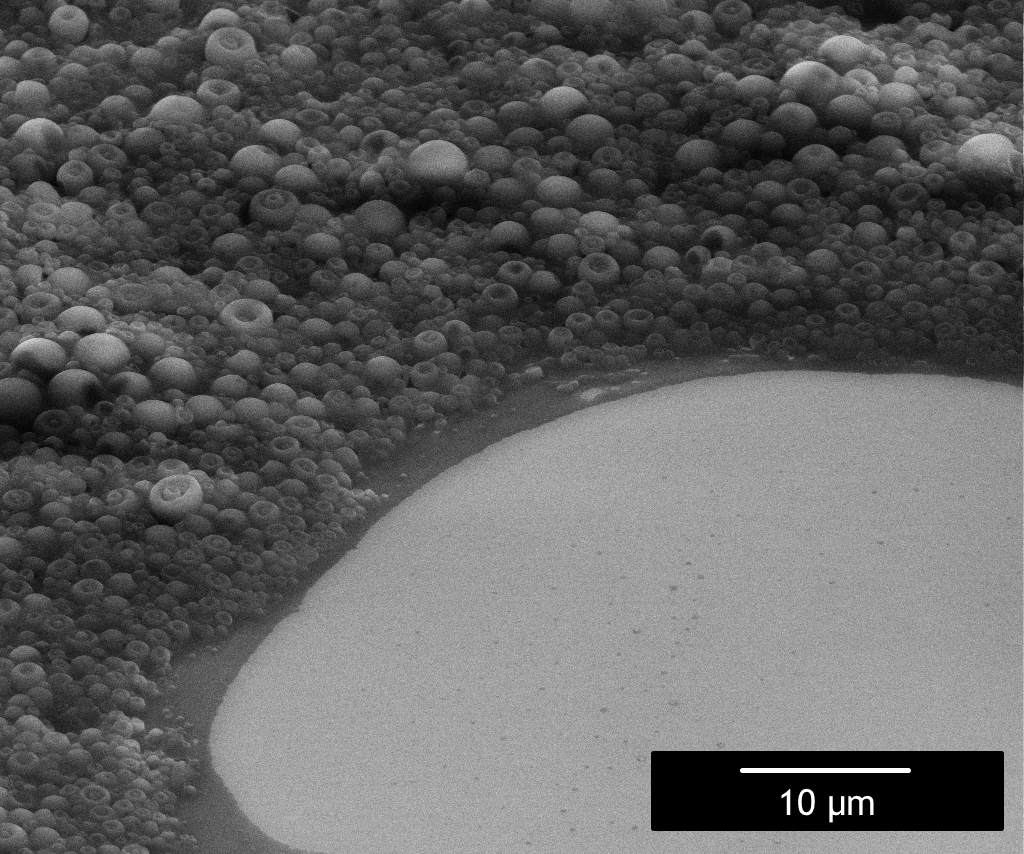
Frixion pen is an erasable ink pen packed with science. This image reveals the microcapsules that contains the thermosensitive ink that becomes invisible when heated.
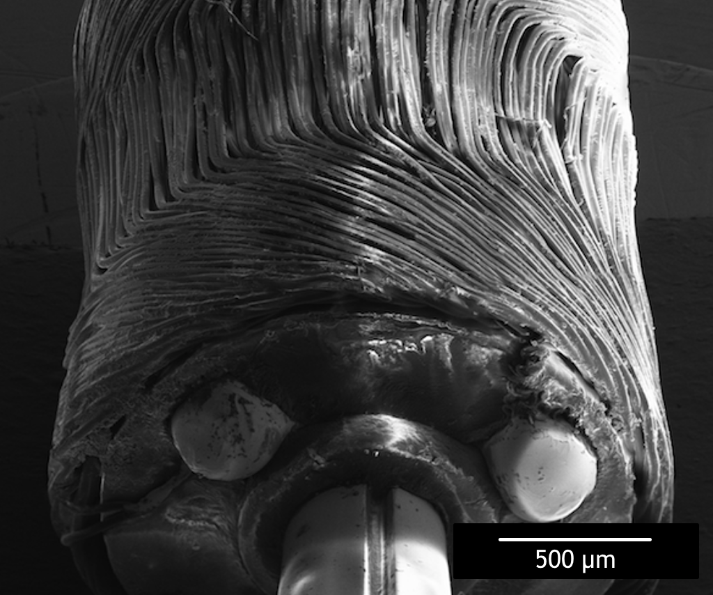
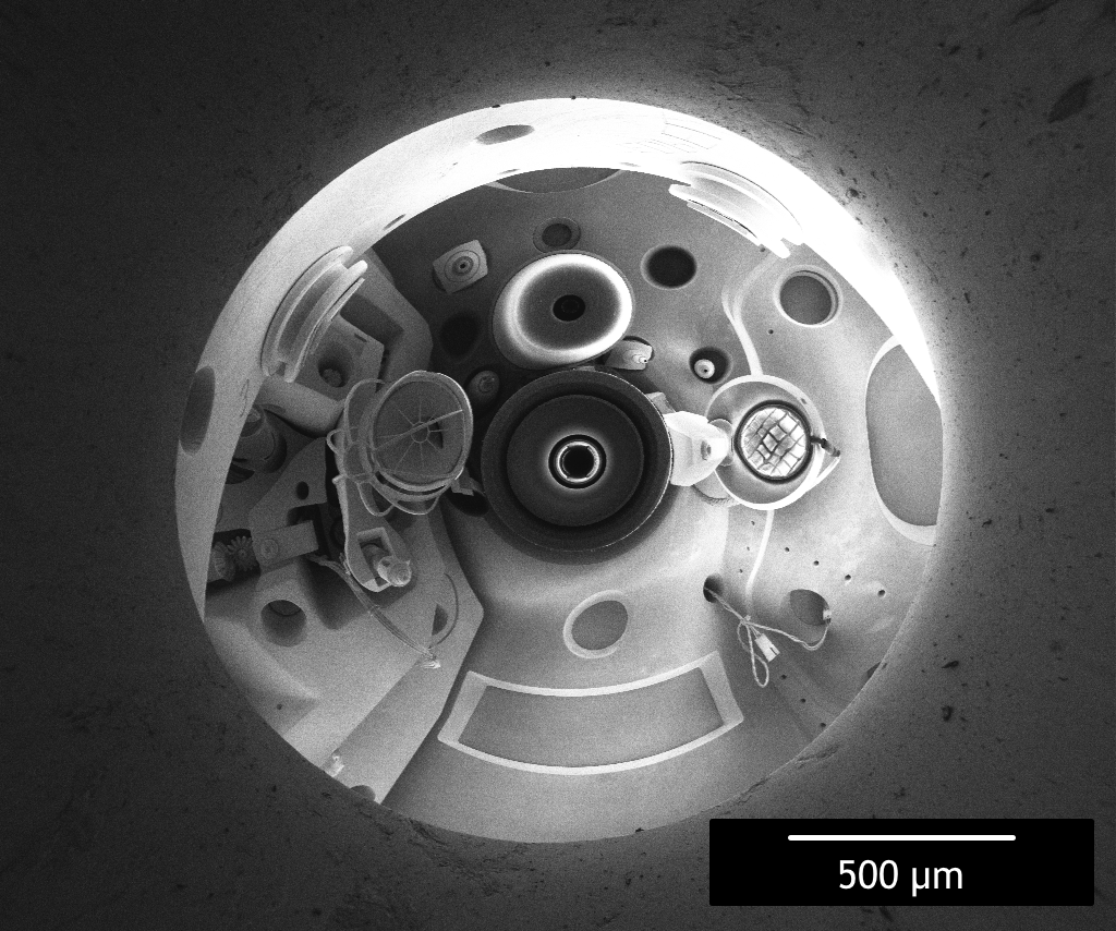
The worlds smallest solar powered car is driven by a microscopic electric motor. This image helps us reverse engineer the motor potentially design a smaller one.
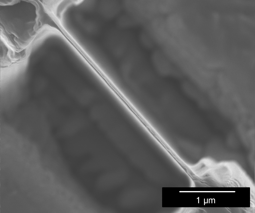
The resolution of a scanning electron microscopy is approximately 5 [nm]. This sample was thinned using a focused ion beam in order to image it in a transmission electron microscope which can resolve atoms.
An electron mirror sample was used to reflect the electron beam to image the objective lens instead of the sample. This technique is used to inspect the electron lens.
This document is the standard operating procedure (SOP) for the FEI DB235 focused ion beam system at UHNF. This SOP serves as a foundation for initial training and ensures that the equipment can be operated correctly, by everyone, the first time.
This is a service manual we developed for the FEI DB235 focused ion beam system at UHNF. This document ensures that any staff can effectively perform routine service or repairs effectively, quickly and at significantly lower cost. This document is restricted to equipment custodians. Contact us for access.